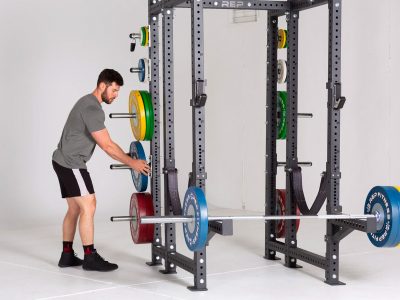
Some diagnostic tests
may be performed to rule out other causes of your pain. These include the
following:
-
An X-ray of the affected area, to
look for bony spurs (abnormal areas) or arthritis;
-
Aspiration, in which fluid is
taken from the swollen bursa and evaluated under a microscope, to rule
out gout or infection;
-
Blood tests to screen for
conditions such as rheumatoid arthritis or diabetes.
A magnetic resonance
imaging test (MRI) also may be performed to evaluate your condition.
What Are the Symptoms of
Bursitis?
-
Pain, inflammation, and swelling
in the shoulders, elbows, hips, knees, or joints of the hands or feet,
particularly during stretching or extension when exercising, lifting,
or otherwise pushing the joint beyond its normal limits;
-
Restricted range of motion in a
joint, with or without immediate pain;
-
Muscle weakness;
-
Loss of motion, especially
affecting the shoulder.
How Is It Treated?
Although bursitis
generally disappears in a few days or weeks, you must take measures to
avoid further strain or injury. A physical therapy program that includes
stretching and focused strengthening exercises could be helpful.
Though you may be
tempted to tough-out the pain, it's not a good idea. Left untreated,
chronic bursitis can lead to the formation of calcium deposits in normally
soft tissues, sometimes causing permanent reduction of motion in the
affected joint.
Initial treatment
typically consists of aspirin or other over-the-counter nonsteroidal
anti-inflammatory drugs (NSAIDs). These pain relievers also tend to reduce
inflammation. An injection of a local anesthetic mixed with one or more
cortisone preparations into the bursa may be recommended to decrease
inflammation and reduce pain. Heat and ultrasound can help relax the joint
and promote tissue repair.
Diathermy (deep-heat
therapy), under the direction of a sports physician, licensed physical
therapist, or trainer, not only can relieve the discomfort and
inflammation of bursitis but also can soothe tense muscles, nerves, and
tendons. It may help to apply ice to the affected area for 20 to 30
minutes twice daily for two to three days.
Your
doctor may prescribe physical therapy to strengthen the
joint and prevent recurrence. Medication may also be
prescribed including anti-inflammatory/ analgesic
medication to relieve pain. When you return to
your sport, go back slowly. Warm up well and do
range-of-motion exercises.
Bursitis may recur,
particularly if you engage regularly in strenuous exercise or physical
labor. In such cases, your doctor may prescribe corticosteroid treatment,
in the form of either oral medication, topical creams, or injections to
the affected joints. In severe cases, it may be necessary to draw fluid
from inflamed and swollen bursae with a needle to relieve the pressure. In
persistent conditions, bursae can be surgically removed.
Red &
Infrared Light Therapy (sometimes called Anodyne treatment)
have proven to increase the blood flow thus providing more
nutrients and oxygen and speed the healing process. This
process in quickly gaining converts and has become accepted
in most hospitals
and PT clinics.
How Can I Prevent
It?
Warming up before
strenuous exercise and cooling down afterward is the most effective way to
avoid bursitis and other strains affecting the bones, muscles and
ligaments. Avoid activities that aggravate the problem. Rest the affected
area after activity.
Call Your Doctor If:
-
Pain in a joint persists more
than a few days; you may be experiencing tendonitis, a strained
ligament or tendon, or the onset of arthritis;
-
You are unable to move the
affected area;
-
Swelling persists after taking a
painkiller or anti-inflammatory agent as prescribed. You may need to
have a doctor drain fluid from the affected joint, or you may need
steroid injections into the joint to decrease inflammation.
-
You have a fever with your
bursitis symptoms.
Some Common Types of
Bursitis
Subacromial
bursitis
The subacromial bursa
lies just above the rotator cuff. Bursitis often develops
due to injury, impingement (pinching), overuse of the
shoulder, or calcium deposits. Symptoms include pain in the
upper shoulder or upper third of the arm, and severe pain
upon moving the shoulder.
Trochanteric
bursitis
The trochanteric bursa
is located over the prominent bone on the side of the hip.
Women and middle-aged to older people are more often
affected by this type of bursitis. It may occur
spontaneously without specific injury. Also, it can be
caused by walking abnormally due to arthritis in the hip,
knee, ankle, foot, or back. Symptoms include pain gradually
occurring over the side of the hip (and sometimes traveling
down the thigh); pain when sleeping on the side affected by
bursitis, rising from a deep chair, sitting in a car, or
climbing stairs; and occasionally, pain when walking.
Ischial
bursitis
The ischial bursa is
located below the bone in your buttock called the ischium.
Inflammation may occur as a result of injury or prolonged
sitting on hard surfaces. Symptoms include pain when sitting
or lying down on the buttocks and pain that travels to the
back of the thigh. Ischial bursitis is also called
"weaver's bottom" or "tailor's seat"
because traditionally, it was an occupational injury of
weaving where there was pressure on the buttocks at the same
time as frequent extension of the hip.
Olecranon
bursitis (Elbow)
Elbow bursitis is caused by the inflammation
of the olecranon bursa located between the skin and bones of
the elbow. Elbow bursitis can be caused by injury or
constant pressure on the elbow (for example, when leaning on
a hard surface). Swelling of this small sac may be also be caused by injury, gout, rheumatoid arthritis,
or infection.
Prepatellar
bursitis (Knee)
The prepatellar bursa
is located beneath the skin and in front of the kneecap. It
becomes inflamed as a result of infection, injury, gout, or
repeated irritation from kneeling. Symptoms include swelling
in the front of the knee that may be painful. Redness and/or
warmth may occur with infection or gout. Infrapatellar
bursitis (clergyman's knee), a similar condition, affects
the infrapatellar bursa, located just below the kneecap.
Pes
anserinus bursitis
Bursitis in the knee
is also called goosefoot bursitis or Pes Anserine bursitis.
The Pes Anserine bursa is located between the shin bone and
the three tendons of the hamstring muscles, on the inside of
the knee. This type of bursitis may be caused by lack of
stretching before exercise, tight hamstring muscles, being
overweight, arthritis, or out-turning of the knee or lower
leg.
Retrocalcaneal
bursitis
This bursa is located
at the back of the heel. Bursitis in this area is often
associated with conditions such as ankylosing spondylitis or
rheumatoid arthritis. It can occur in healthy individuals
who wear improperly fitted shoes. Symptoms include painful
swelling that develops at the back of the heel.
Calcaneal
bursitis
This bursa is located
at the sole or bottom of the heel. Inflammation usually
produces pain in the heel when standing. Causes include heel
spurs, excess weight, injury, and wearing improperly fitted
shoes.
Anterior Achilles
tendon bursitis
This type of bursitis
is also called Albert's disease. Extra strain on the
Achilles tendon, such as injury, disease, or shoes with
rigid back support, causes this condition, which is
characterized by inflammation of the bursa located in front
of the attachment of the tendon to the heel.
Posterior Achilles Tendon
Bursitis
This type of bursitis,
also called Haglund's deformity, is located between the skin
of the heel and the Achilles tendon (which attaches the calf
muscles to the heel). Aggravated by a type of walking that
presses the soft heel tissue to the hard back support of a
shoe, this type of bursitis occurs mostly in young women.
See: Achilles
Tendon - Bursitis
We
gratefully acknowledge this as a contribution of WebMD